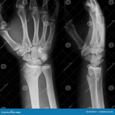
X-ray image of Left wrist joint AP and Lateral view for showing fracture of radius bone Stock ... is a high-quality image in the Cleve collection, available at 1034 × 1390 pixels resolution — ideal for both digital and print use.
Understand the purpose and procedure of a wrist X-ray. Our guide explains how this diagnostic imaging tool identifies fractures, dislocations, arthritis, and bone injuries. Learn what to expect during your orthopedic exam, how radiologists interpret these medical images, and why this non-invasive test is essential for evaluating wrist pain and ensuring proper bone health recovery.
Image Details
| Title | X-ray image of Left wrist joint AP and Lateral view for showing fracture of radius bone Stock ... |
|---|---|
| Dimensions | 1034 × 1390 px |
| Category | Cleve |
| Published | March 27, 2026 |
| Author | Zeus |
| Downloads | 2,089 |
| Views | 129 |
Read full article: Wrist X Ray