Knee Anatomy: Muscles, Ligaments, and Cartilage - JOI Jacksonville Orthopaedic Institute is a high-quality image in the Cleve collection, available at 1024 × 1024 pixels resolution — ideal for both digital and print use.
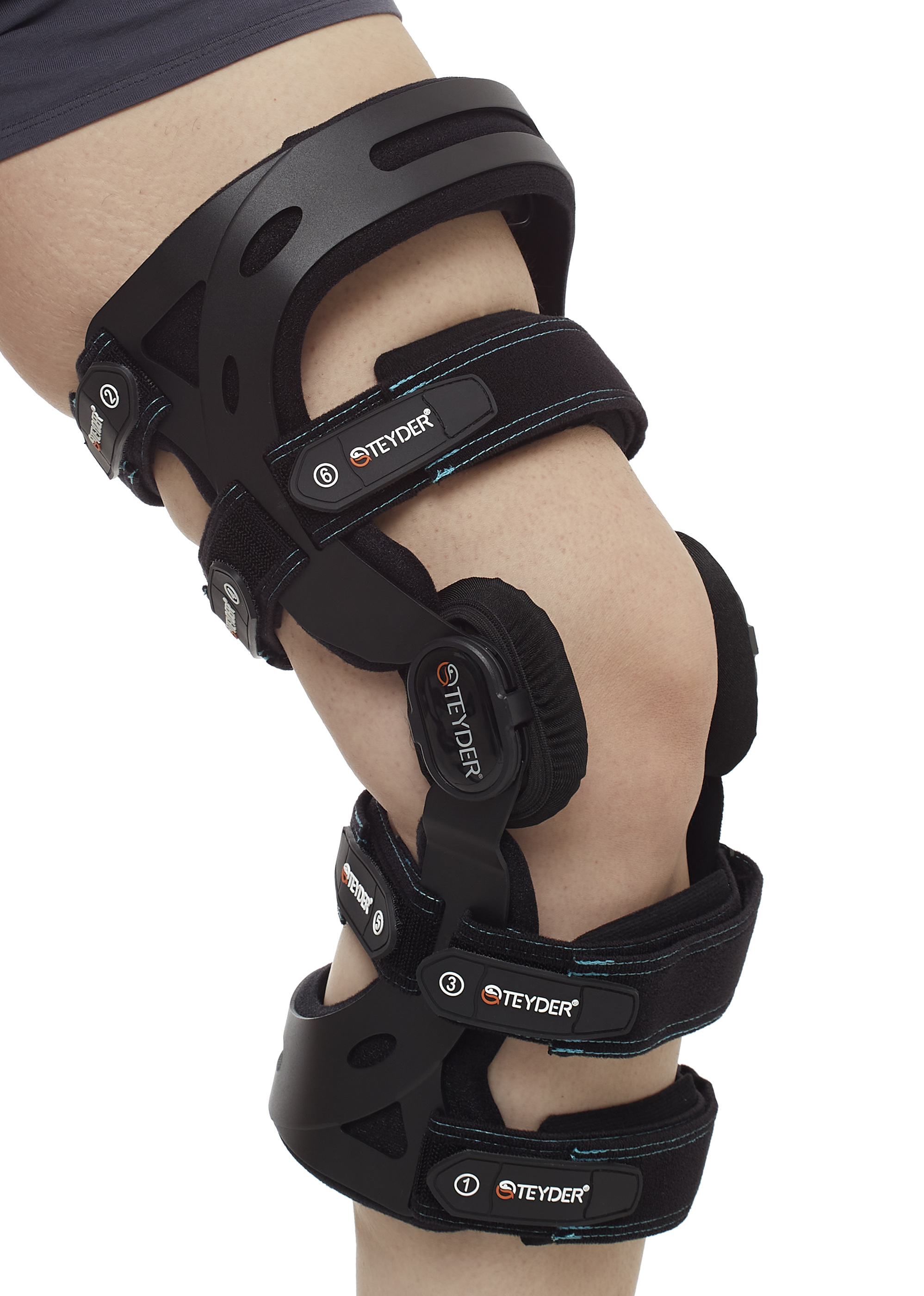
Explore the complex knee anatomy ligaments in detail. Learn how the ACL, PCL, MCL, and LCL provide structural support and joint stability. Our comprehensive guide explains their vital roles in movement, common injury patterns, and essential knee health, helping you understand how these connective tissues keep your legs functioning properly and pain-free during daily activities and sports.
Image Details
| Title | Knee Anatomy: Muscles, Ligaments, and Cartilage - JOI Jacksonville Orthopaedic Institute |
|---|---|
| Dimensions | 1024 × 1024 px |
| Category | Cleve |
| Published | March 14, 2025 |
| Author | Zeus |
| Downloads | 1,189 |
| Views | 1,232 |
Read full article: Knee Anatomy Ligaments