Histological Sample Simple Columnar Epithelium Tissue Under the Microscope. Stock Photo - Image ... is a high-quality image in the Cleve collection, available at 1600 × 1157 pixels resolution — ideal for both digital and print use.
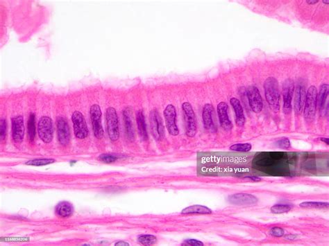
Discover how to observe epithelial cells under a microscope with this detailed guide. Learn essential staining techniques, proper slide preparation, and how to identify cellular structures like the nucleus and cell membrane. Master microscopy basics to visualize tissue layers, morphology, and specimen characteristics clearly for your biology studies or laboratory research.
Image Details
| Title | Histological Sample Simple Columnar Epithelium Tissue Under the Microscope. Stock Photo - Image ... |
|---|---|
| Dimensions | 1600 × 1157 px |
| Category | Cleve |
| Published | March 19, 2026 |
| Author | Zeus |
| Downloads | 777 |
| Views | 1,238 |
Read full article: Epithelial Cells Under Microscope