RADIOGRAPHIC ANATOMY OF KNEE JOINT AND ITS RADIOGRAPHIC VIEWS.pptx is a high-quality image in the Cleve collection, available at 2048 × 1152 pixels resolution — ideal for both digital and print use.
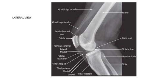
Discover the intricate bone anatomy of the knee. Learn how the femur, tibia, patella, and fibula work together to support movement and stability. This comprehensive guide breaks down key skeletal structures, joint mechanics, and cartilage function, helping you understand how these vital components contribute to overall leg health and common orthopedic conditions.
Image Details
| Title | RADIOGRAPHIC ANATOMY OF KNEE JOINT AND ITS RADIOGRAPHIC VIEWS.pptx |
|---|---|
| Dimensions | 2048 × 1152 px |
| Category | Cleve |
| Published | April 4, 2026 |
| Author | Zeus |
| Downloads | 1,635 |
| Views | 890 |
Read full article: Bone Anatomy Knee