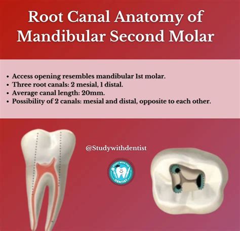
Root Anatomy And Root Canal Morphology Of Maxillary Second – ZCPI is a high-quality image in the Rp collection, available at 1062 × 1026 pixels resolution — ideal for both digital and print use.
Explore the complex anatomy of the mandibular bone, including its essential structure, mental foramen, and alveolar process. This guide details how these anatomical landmarks relate to dental function, nerve pathways, and jaw health. Learn the clinical significance of mandibular morphology and how it impacts oral surgery and orthodontic treatments in this comprehensive breakdown of human jaw physiology.
Image Details
| Title | Root Anatomy And Root Canal Morphology Of Maxillary Second – ZCPI |
|---|---|
| Dimensions | 1062 × 1026 px |
| Category | Rp |
| Published | January 13, 2026 |
| Author | Zeus |
| Downloads | 2,258 |
| Views | 1,309 |
Read full article: Anatomy Of Mandibular